Dry eye fluoresceins and acinar-meibography
The EyePhotoDoc’s LED exciter and barrier filter show more detail
A photograph is more descriptive than words. A video shows tear dynamics. Scientific explanation below
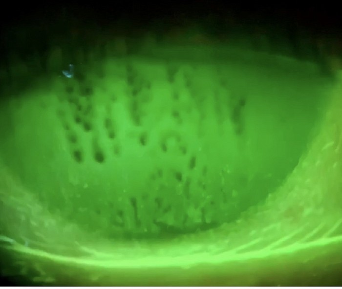
Subtle punctate changes maybe missed
Most significant way to help your dry eye patients
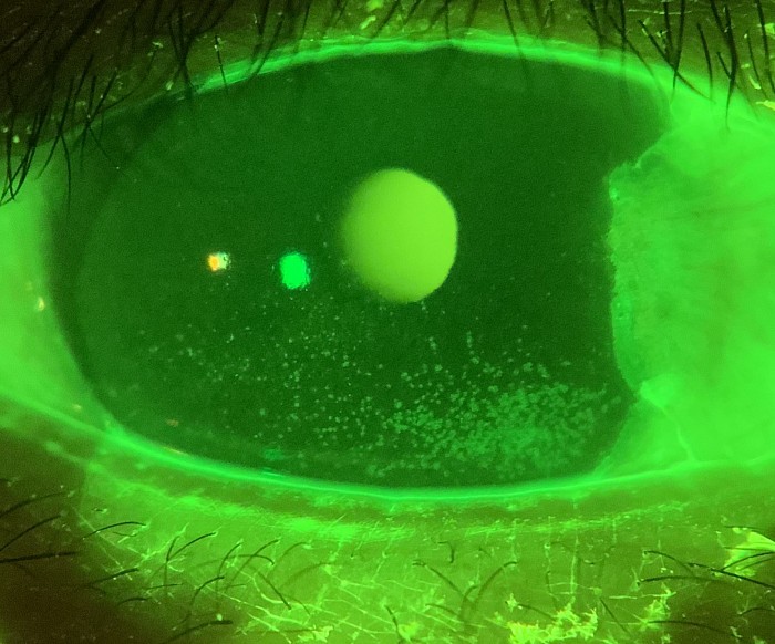
Office tests for dry eye diagnosis have many non-conclusive tests. Unfortunately they are only suggestive such as the Schrimer test, TBUT, tear osmolarity, but are not diagnostic. Our advanced fluorescent Diode and yellow barrier filter find subtle punctate changes. Punctate stain is the only objective test for dry eye. It also tells the degree and severity that corresponds to patient’s complaints.
Photos help in patient follow up
Evidenced based dry eye treatment
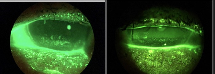
This patient was treated for dry eyes. The left represents the original findings while the right shows the improvement a month later. Note the change in punctuate stain and the reestablishment of the tear film.
Below is an important new development. Watch the video of the blink and tear film dynamics. Note the tear film coating, it’s spread and breakup. This is called dynamic tear film analysis.
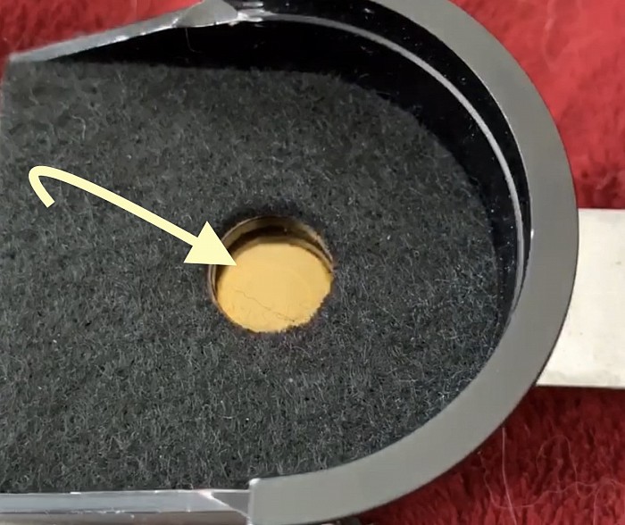
Slide in blue barrier filter
How this works: Blue Barrier filter
The filter slides in and out. This permits external photos and quickly converts to yellow Wratten type filter
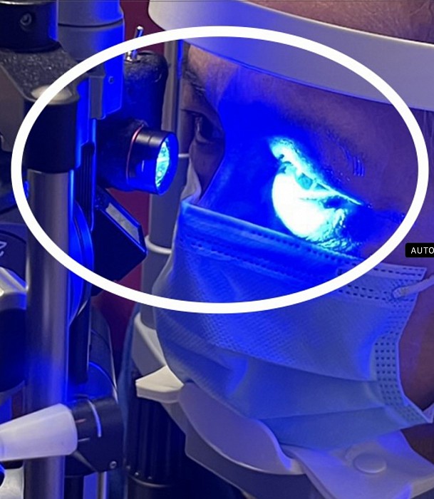
Full field fluorescein LED exciter
Brighter and wider fluorescent diode beam
The special fluorescein exciting LED light is designed especially for diagnosis of external ocular tear film abnormalities. Unlike the typical slit lamp 10 mm blue light it is more intense and has a greater field. It allows you to see the lid action, tear film spread and breakup like never before.
Highly Recommended if your slit lamp does not have a yellow filter
Why this works better:
The standard slit lamp blue light washes out the fluorescent green detail. By using a yellow barrier filter the blue color is blocked and only the fluorescent detail is seen. The tear film and ocular surface disorders are easily seen. Click here for more videos
The legacy ‘tear break up time’ can be videoed for more understanding of the tear film behavior. The images can be used to show the patient and evaluated the efficacy of treatment.
The technology is ideal to evaluate different treatments. The before and after images help you decide on the type of drops, value of Restasis™️ and other meds in the category, punctal plugs, serum drops, meibomian treatments, etc
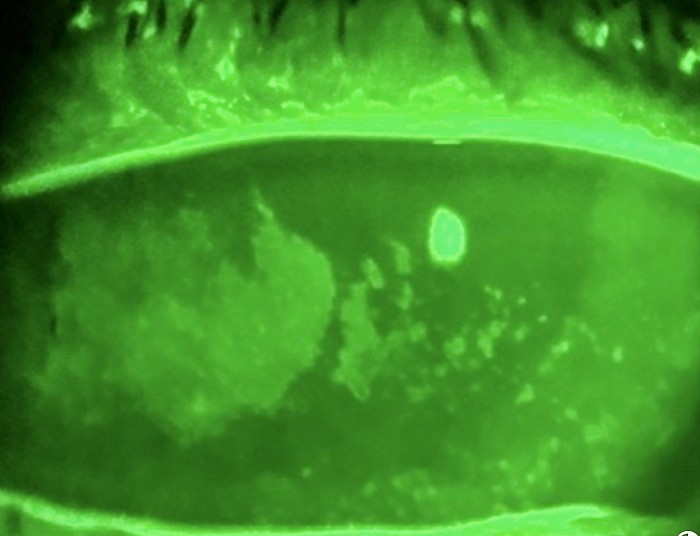
Punctate Stain
Mucus in tears
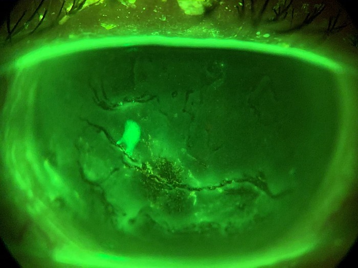
Epithelial breakdown
Now Acinar-Meibography for diagnosis and treatment of oil layer deficiencies and accessory lacrimal gland disorders
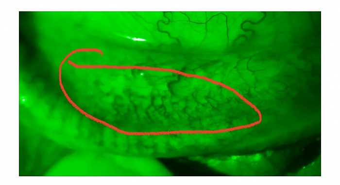
Meibomian glands and accessoryu lacrimal glands
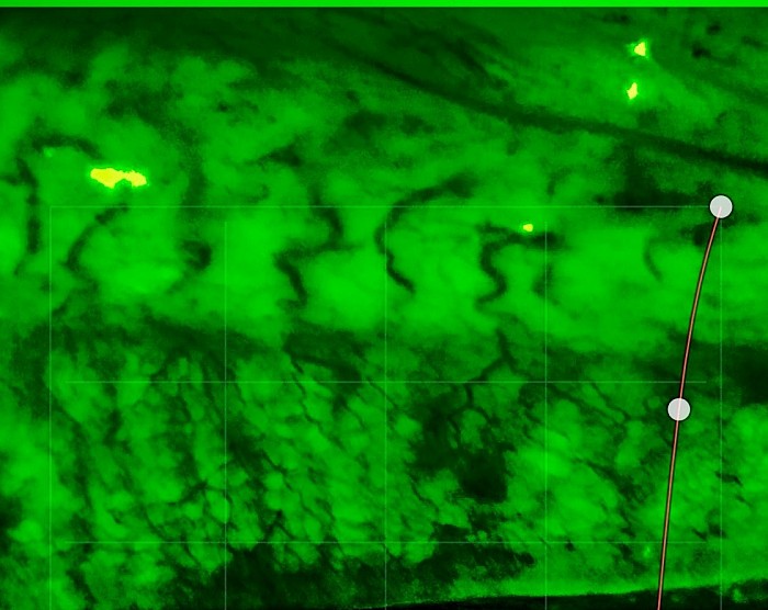
The acinar-meibography red-free light technology is a significant advancement in imaging for dry eye examinations. By using red-free light, it offers improved visualization of the meibomian glands and accessory lacrimal glands, allowing for greater detail and clarity compared to traditional infrared (IR) imaging. The ability to magnify the glands with a slit lamp’s higher power further enhances the level of detail that can be observed.
One notable advantage of this technology is its utility in diagnosing and monitoring acinar-meibography disease. It enables clinicians to identify abnormalities such as orifice plugging, acinar space swelling, loss of acinar structures, and gland drop-out, which are indicative of gland dysfunction. This information is crucial for selecting appropriate therapeutic interventions and monitoring the progression of the disease over time.
Moreover, the integration of the EyePhotoDoc Apple camera enhances documentation and facilitates communication with patients by capturing detailed images of the vascularization of the meibomian glands and accessory glands. This comprehensive approach to imaging and documentation is invaluable for comprehensive dry eye management.
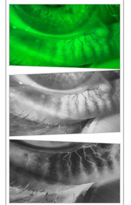
Photographic acinar-meibography with monochromatic and inverse filters
New meibography techniques
By using your camera you get higher resolution as compared too IR meibography cameras. Above is a green photo, middle a monochromatic photo used to accentuate the glands, and below an inverse demonstrating loss of vessels in dying glands.
See the acinar spaces
High power slit lamp observation of meibomian glands
The ability to use magnification on the slit lamp to observe meibomian gland morphology in detail is indeed a valuable tool for assessing dry eye patients. With this level of magnification, clinicians can identify subtle abnormalities in gland structure and function that may contribute to dry eye symptoms. Additionally, being able to document these findings with high-quality photos allows for better tracking of treatment progress and response over time.
By routinely scanning every dry eye patient for meibomian gland pathology, clinicians can proactively identify and address issues that may exacerbate dry eye symptoms. This approach not only helps in individualizing treatment plans but also contributes to better overall management of dry eye disease.
Overall, the combination of slit lamp magnification and documentation with high-quality photos provides clinicians with a comprehensive and effective means of evaluating and managing dry eye patients.
For more information click here
See more informative videos
