Slit lamp Ophthamoscope
Transformational device
Slit lamp opthalmoscope
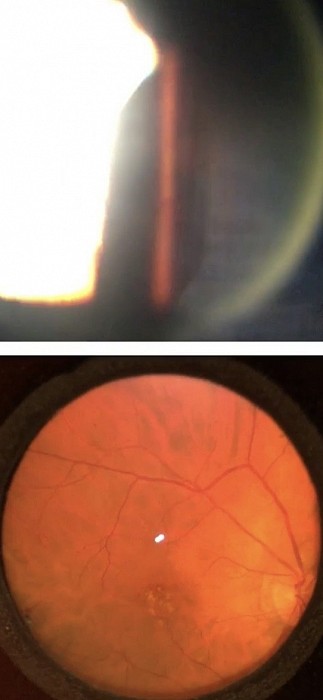
The upper right shows the narrow 5° view of the slit lamp fundoscopy 60D lens.
Below is our new slit lamp ophthalmoscope 45° view of the Fundus. This revolutionary device uses much less light for patient comfort. The wide view takes away the concern of missing findings. Users have compared it to a ‘live’ highly detailed Fundus photo and a magnified indirect ophthalmoscope view.
Globe positioning system
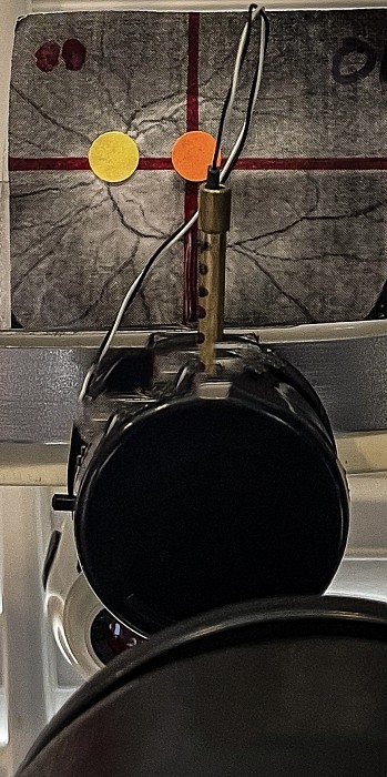
GPS (Globe positioning system)
Always know where you are viewing. No longer following the vessels to find the optic nerve. ‘Look at my ear fixation’ has been improved by a fixation light that goes right and left and up and down. It is calibrated in degrees using macular fixation. To take away any equivocation of retinal location since there is a map of where you are viewing.
Slit Lamp ophthalmoscope
Slit lamp Ophthalmoscope
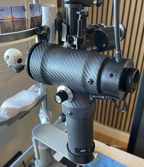
The slit lamp Ophthalmoscope is built from the ground up to image the retina. The fundoscopy lenses were a clever afterthought to work with our slit lamps. Well known to all of us are the shortcomings of these bio lenses. The light is so intense it is bothersome to the patient with the glare and reflections are a nuisance to the doctor. The actual use of the lens is cumbersome to hold.
The slit lamp Ophthalmoscope attaches to the slit lamp to steady the head and the carriage to focus onto the retina. There is a broad field of view with no bothersome glare. The free hand works the GPS to view different locations in the retina. The map indicates where you are viewing.
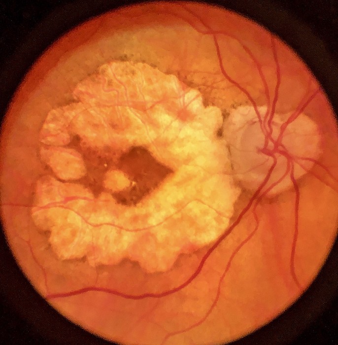
Below are some photos taken with the slit lamp Ophthalmoscope. After decades of clinical practice I can attest how much easier it is to visualize the entire picture of what is going on with a large magnified field in comparison to a slit! Note the photos are less detailed than your view in the ophthalmoscope.
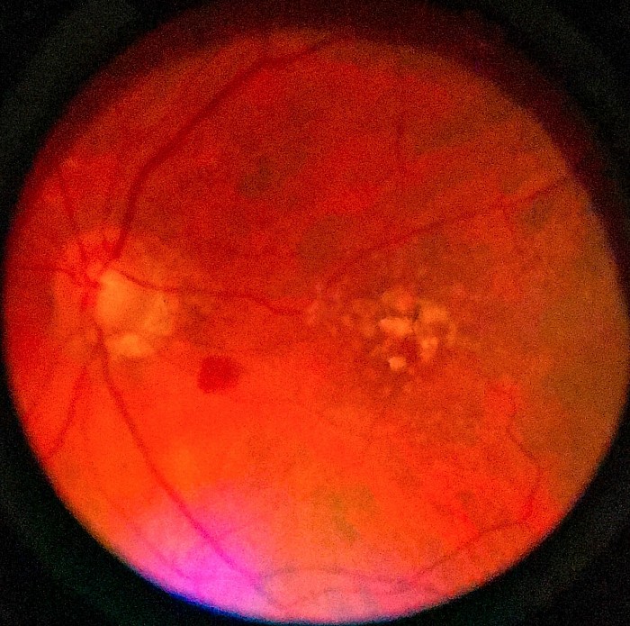
Macular Degeneration
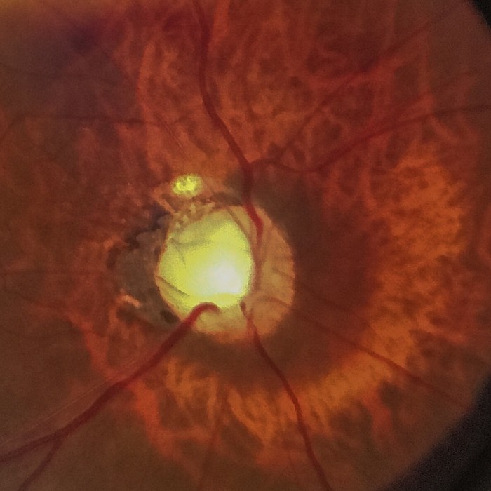
Optic nerve atrophy
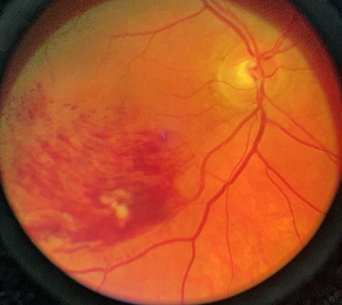
Branch vein occlusion
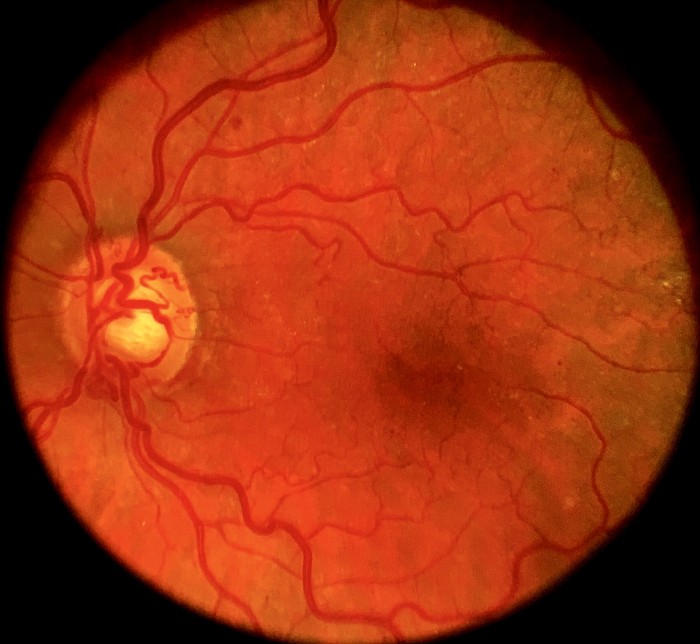
Vasculitis
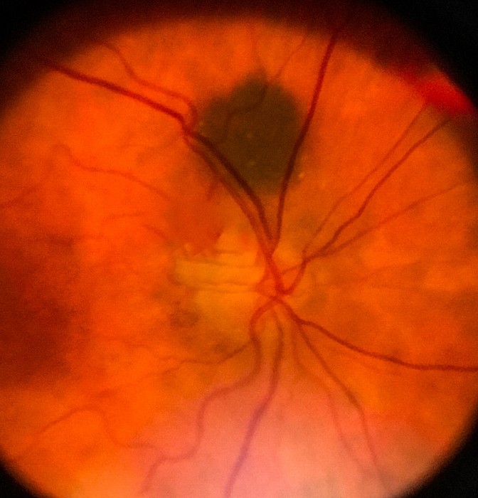
Nevus
Choroidal atrophy
Availability in March
We have designated 5 units for teaching institutes free of cost. The early adopter units will be available for $1500 to eye doctors. Contact Dr. Terry at info@eyephotodoc.com for more information.
